Which part of the heart forms the lateral border
On the lateral projection the cardiac silhouette is formed by 1: the anterior border by right ventricle. the posterior border by left atrium (superiorly) and left ventricle (inferiorly) and the inferior vena cava.
What is the lateral part of the heart?
Lateral Wall | Atlas of Human Cardiac Anatomy. Location: The lateral wall is generally considered to include the wall of the right atrium from the ostia of the superior and inferior vena cava anteriorly to the ostium of the right appendage or auricle.
What forms the left border of the heart?
Anatomical terminology The left border of heart (or left margin, or obtuse margin) is shorter than the right border, full, and rounded: it is formed mainly by the left ventricle, but to a slight extent, above, by the left atrium.
What part of the heart forms the right border?
The right border of the heart extends just over 1 cm to the right of the sternum, between the 3rd and 6th intercostal cartilages. It is composed mainly of the right atrium. The inferior border runs from the 6th costal cartilage on the right, through the xiphisternal joint, to the 5th intercostal space on the left.What are the borders of the heart?
- right border: IVC, right atrium, SVC.
- left border: left ventricle, left atrium, pulmonary trunk and arch of aorta.
- inferior border: right ventricle.
- superior border: right and left atria, SVC, ascending aorta and pulmonary trunk.
What divides the left and right side of the heart?
S. septum (SEP-tum): The septum is a thick wall of muscle that divides the heart. It separates the left and right sides of the heart.
What is proximal to the heart?
If another reference point is given, such as the heart, the proximal point of another organ or extremity is the point closest to the heart, central rather than peripheral. … Proximal is the opposite of distal.
Where is upper border of the heart?
The upper border is hidden behind the sternum at the level of the second and third cartilages. The right margin of the heart peeps out behind the right border of the sternum between the right third and sixth cartilages.What part of the heart forms the diaphragmatic surface?
The diaphragmatic surface (inferior surface), directed downward and slightly backward, is formed by the ventricles, and rests upon the central tendon and a small part of the left muscular portion of the diaphragm.
Is the heart surrounded by cartilaginous pericardium?The pericardium, which literally translates as “around the heart,” consists of two distinct sublayers: the sturdy outer fibrous pericardium and the inner serous pericardium. The fibrous pericardium is made of tough, dense connective tissue that protects the heart and maintains its position in the thorax.
Article first time published onHow do you find the left border of the heart?
The left border of the relative cardiac dullness is normally found in the 5th intercostal space 1.5 cm medially of the left midclavicular line. Displacement of the relative cardiac dullness borders may occur due to dilation of the heart chambers and to a lesser extent due to thickening (hypertrophy) of myocardial wall.
Where is base of the heart?
The base of the heart is located at the level of the third costal cartilage, as seen in Figure 1. The inferior tip of the heart, the apex, lies just to the left of the sternum between the junction of the fourth and fifth ribs near their articulation with the costal cartilages.
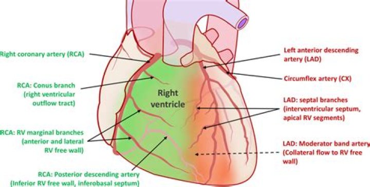
What is the RCA in the heart?
Right coronary artery (RCA). The right coronary artery supplies blood to the right ventricle, the right atrium, and the SA (sinoatrial) and AV (atrioventricular) nodes, which regulate the heart rhythm.
Where is the base of the heart located quizlet?
The heart is located between the sternum and the vertebral column. About two thirds of the heart’s mass lies to the left of the body’s midline. The base of the heart rests on the diaphragm.
What are the borders in anatomy?
[bor´der] a bounding line, edge, or surface. brush border a specialization of the free surface of a cell, consisting of minute cylindrical processes (microvilli) that greatly increase the surface area. Brush border, characterized by closely packed microvilli.
What are the anatomical relations of the heart?
The heart is made up of four chambers: two upper chambers known as the left atrium and right atrium and two lower chambers called the left and right ventricles. It is also made up of four valves: the tricuspid, pulmonary, mitral and aortic valves.
Which side is medial lateral?
A lateral orientation is a position away from the midline of the body. For instance, the arms are lateral to the chest, and the ears are lateral to the head. A medial orientation is a position toward the midline of the body. An example of medial orientation is the eyes, which are medial to the ears on the head.
Is the heart lateral to the sternum?
Towards the back of the body. The heart lies posterior the sternum. You just studied 24 terms!
Are the shoulders lateral or medial to the midline?
The neck is medial to the shoulder. Lateral: Farther from the midline.
What are the downstairs chambers called?
The upper chamber is called an atrium (or auricle), and the lower chamber is called a ventricle. The two atria act as receiving chambers for blood entering the heart; the more muscular ventricles pump the blood out of the heart.
Where is Vena Cava?
The inferior vena cava (IVC) is the largest vein of the human body. It is located at the posterior abdominal wall on the right side of the aorta. The IVC’s function is to carry the venous blood from the lower limbs and abdominopelvic region to the heart.
Where is the septum on the heart?
Heart septum: The dividing wall between the right and left sides of the heart.
Is the heart anterior to the vertebral column?
The heart is situated in the middle of the two lungs and in front of a vertebral column in a thoracic cavity. However it is located posterior(behind) to the breastbone plate i.e sternum.
How many borders does the heart have?
The heart has four borders: right border: IVC, right atrium, SVC. left border: left ventricle, left atrium, pulmonary trunk and arch of aorta. inferior border: right ventricle.
Where is the second intercostal space?
From the angle of Louis, move your fingers to the right and you will feel a gap between the ribs. This gap is the 2nd Intercostal space. From this position, run your fingers downward across the next rib, and the next one.
Is the pericardium the outer layer of the heart?
The Pericardium. The pericardium is the fibrous sac that surrounds the heart. … The inner surface of the fibrous pericardium is lined by the outer (parietal) layer of serous pericardium. The inner (visceral) layer of the serous pericardium lines the outer surface of the heart itself.
Which layer is both a part of the pericardium and a part of the heart wall?
The visceral layer of the serous pericardium (epicardium) is both a part of the pericardium and a part of the heart wall.
What is the outer layer of the pericardium?
PericardiumLocationA sac around the heartArteryPericardiacophrenic arteryNervePhrenic nerveIdentifiers
What forms the right and left heart border in a chest xray?
On a PA view, the right heart border is formed by the right atrium, and the left heart border is formed by the appendage of the left atrium superiorly and the left ventricle inferiorly.
Where does the left ventricle sit in the chest?
1. Location of the heart in the center of the chest. The figure shows a transverse section. The left ventricle actually lies behind and to the left of the right ventricle.
Why is it called the base of the heart?
The base of the heart is probably better termed its posterior surface. … It assumed the term because it is thought to resemble the base of the pyramid or cone which extends obliquely to the left to the apex of the heart. The surface of the base is quadrangular in shape and faces posteriorly and slightly to the right.