Where is the internal venous plexus located
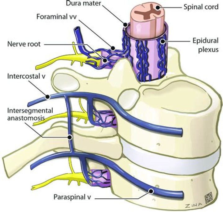
The internal vertebral venous plexus is located beneath the bony elements of the vertebral foramina (laminae, spinous processes, pedicles, and vertebral body). It is embedded in a layer of loose areolar tissue know as the epidural (extradural) adipose tissue.
What are venous plexuses?
A venous plexus is a congregation of multiple veins.
Where does main part of vertebral venous plexus lies?
Median sagittal section of two thoracic vertebrae, showing the vertebral venous plexuses. The internal vertebral venous plexuses (intraspinal veins) lie within the vertebral canal in the epidural space, and receive tributaries from the bones and from the spinal cord.
Where does the internal venous plexus drain?
The internal plexus drains into the external one, which in turn drains into the vertebral veins of the neck and segmental (intercostal, lumbar, and sacral) veins of the trunk.What is Batson venous plexus?
Batson venous plexus, also known as Batson veins, are a network of paravertebral veins with no valves that connect thoracic vessels and deep pelvic veins draining the bladder, prostate, and rectum to the internal vertebral venous plexus 1.
What does the internal jugular vein drain?
The function of the internal jugular vein is to collect blood from the skull, brain, superficial parts of the face, and the majority of the neck. … The blood collected from these vessels then drains to the brachiocephalic vein and into the right atrium.
Where does prostatic venous plexus drain?
The receipt of blood from the vesical and prostatic rami connect the prostatic plexus to the vesical plexus and internal pudendal vein. The prostatic plexus then drains into the vesical and internal iliac veins.
What is portal venous system?
In the circulatory system of animals, a portal venous system occurs when a capillary bed pools into another capillary bed through veins, without first going through the heart. Both capillary beds and the blood vessels that connect them are considered part of the portal venous system.What drains into pterygoid plexus?
In addition, the inferior ophthalmic vein and deep facial vein also drain into the pterygoid plexus. The plexus itself drains via the short maxillary vein before it forms the retromandibular vein. Emissary veins also anastomose between the plexus and the cavernous sinus, via the foramina ovale and lacerum.
What is vertebral venous plexus?The vertebral venous plexus is generally described as a thin walled, valveless net- work of veins within and surrounding the vertebral column and extending from. cranium to pelvis. It consists of an internal division which lies within the epidural. space with direct connections to the vertebral bodies.
Article first time published onWhat is vertebral plexus?
The vertebral venous plexus, also called the Batson plexus refers to veins located extradurally within the spinal canal (internal plexus), surrounding the vertebral column (external plexus) as well as horizontal basivertebral veins that run through the vertebral bodies (basivertebral plexus).[1][2] These veins comprise …
What does the internal thoracic vein drain into?
The internal thoracic vein arises from the superior epigastric vein. … After uniting, each internal thoracic vein drains into its respective brachiocephalic vein 2.
What drains into Batson's plexus?
The Batson venous plexus (Batson veins) is a network of valveless veins in the human body that connect the deep pelvic veins and thoracic veins (draining the inferior end of the urinary bladder, breast and prostate) to the internal vertebral venous plexuses.
What is the clinical significance of the spinal valveless plexus?
Today, the VVP is considered part of the cerebrospinal venous system, which is regarded as a unique, large-capacitance, valveless plexiform venous network in which flow is bidirectional that plays an important role in the regulation of intracranial pressure with changes in posture and in venous outflow from the brain, …
How does the Pampiniform plexus regulate temperature?
The pampiniform plexus helps regulate the temperature of the testes by acting as a “heat exchange” mechanism to cool down the blood. The arteries supplying the testes run through the plexus where the blood is cooled from abdominal arterial temperature to testicular temperature.
What is the prostatic plexus?
The prostatic plexus is a relatively large bundle of nerves that arises from the inferior (lower) portion of the pelvic plexus, a bundle of nerves, located on either side of the rectum. It is located in the prostate’s fascial shell, a layer of connective tissue.
Where does internal jugular vein start?
The internal jugular vein originates within the posterior part of the jugular foramen, under the posterior part of the floor of the tympanic cavity. The IJV is continuous with the sigmoid sinus; however, its origin is demarcated by a dilation called the superior bulb of internal jugular vein.
Where does the internal jugular vein originate?
The internal jugular vein originates at the jugular foramen and ends up behind the sternoclavicular joint where it joins the subclavian vein. It lies right alongside the carotid artery and the vagus nerve within the carotid sheath.
What vein leads to the internal jugular vein?
Internal jugular veinDrains tobrachiocephalic veinArteryinternal carotid, common carotidIdentifiersLatinvena jugularis interna
Where is the pterygoid plate located?
Two pairs of bony plates, the pterygoid processes, arise from the base of each alisphenoid bone. The outer plates are nearly horizontal in position. They extend from the posterior end of the maxillary bone caudad and laterad to the lateral surface of the tympanic bulla.
Which vein does not join the pterygoid plexus of veins?
The pterygoid plexus of veins becomes the maxillary vein. The maxillary vein and the superficial temporal vein later join to become the retromandibular vein.
How is cavernous sinus related to pterygoid plexus?
The pterygoid plexus communicates with the cavernous sinus through the foramina ovale, spinosum, and rotundum. Blood also leaves the superior and inferior ophthalmic veins to enter first the anterior facial vein, then goes to a separate channel that connects the internal and external jugular veins.
What side is the portal vein on?
Measuring approximately 8 cm (3 inches) long in adults, the portal vein is located in the right upper quadrant of the abdomen, originating behind the neck of the pancreas. In most individuals, the portal vein is formed by the union of the superior mesenteric vein and the splenic vein.
Where is the hepatic portal system located?
Located in the right upper quadrant of the abdomen, it consists of two major lobes that are closely associated with the inferior vena cava. Venous blood rich in nutrients enters the liver from the hepatic portal venous system.
Where are the portal systems in the body?
circulatory system Lower vertebrates have two so-called portal systems, areas of the venous system that begin in capillaries in tissues and join to form veins, which divide to produce another capillary network en route to the heart. They are called the hepatic (liver) and renal (kidneys) portal systems.
Which vein connects the sigmoid sinus to the external venous vertebral plexus?
The posterior and lateral condylar veins allow for connections with the external vertebral venous system, whereas the anterior condylar veins are predominantly related to the internal vertebral venous plexus.
What is a plexus?
A plexus is a bundle of intersecting nerves, blood vessels, or lymphatic vessels in the human body. These bundles typically originate from the same anatomical area and serve specific areas of the body. Bundles of nerves that form a plexus communicate information to your brain about pain, temperature, and pressure.
What is cervical plexus?
The cervical plexus is a network of nerve fibres that supplies innervation to some of the structures in the neck and trunk. It is located in the posterior triangle of the neck, halfway up the sternocleidomastoid muscle, and within the prevertebral layer of cervical fascia.
Where does the internal thoracic artery come from?
To recap, the internal thoracic artery originates off of the subclavian artery giving off several branches as it descends along the inner surface of the anterior thorax: Anterior intercostal arteries at each intercostal space.
Where does the left superior intercostal vein drain into?
The left superior intercostal vein drains the 2nd and 3rd posterior intercostal veins on the left side of the body. It usually drains into the left brachiocephalic vein. It may also communicate with the accessory hemiazygos vein.
Where do anterior intercostal veins drain into?
The anterior intercostal veins originate from the intercostal space just inferior to anterior aspects of their respective ribs and drain into the internal thoracic and musculophrenic veins.