What structures are found in gram negative bacteria?
What structures are found in gram negative bacteria?
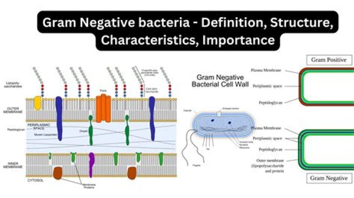
Gram-negative bacteria are surrounded by a thin peptidoglycan cell wall, which itself is surrounded by an outer membrane containing lipopolysaccharide. Gram-positive bacteria lack an outer membrane but are surrounded by layers of peptidoglycan many times thicker than is found in the Gram-negatives.
What is the shape of gram negative bacteria?
In addition to spherical or rod-shaped, Gram-negative bacteria can also be spiral-shaped (spirochetes). Gram-negative bacteria account for a multitude of conditions, including many foodborne illnesses, cholera, gonorrhea, and urinary tract infections.
Is peptidoglycan found in Gram-negative?
Peptidoglycan serves a structural role in the bacterial cell wall, giving structural strength, as well as counteracting the osmotic pressure of the cytoplasm. The peptidoglycan layer is substantially thicker in Gram-positive bacteria (20 to 80 nanometers) than in Gram-negative bacteria (7 to 8 nanometers).
What is significant about Gram negative bacteria?
Gram-negative bacteria are resistant to multiple drugs and are increasingly resistant to most available antibiotics. These bacteria have built-in abilities to find new ways to be resistant and can pass along genetic materials that allow other bacteria to become drug-resistant as well.
What is a Gram-negative organism how does the structure of a Gram-negative organism contribute to its virulence?
The outer membrane of Gram-negative bacteria contains lipopolysaccharides, proteins, and phospholipids. The lipopolysaccharide component acts as a virulence factor and causes disease in animals. More virulence factors are harbored in the periplasmic space between the outer membrane and the plasma membrane.
What is the structure of cell wall in Gram-negative bacteria?
The Gram-negative cell wall is composed of a thin, inner layer of peptidoglycan and an outer membrane consisting of molecules of phospholipids, lipopolysaccharides (LPS), lipoproteins and sutface proteins. The lipopolysaccharide consists of lipid A and O polysaccharide.
What type of cells have peptidoglycan as part of the cell wall?
Peptidoglycan. Unique features of almost all prokaryotic cells (except for Halobacterium halobium and mycoplasmas) are cell wall peptidoglycan and the specific enzymes involved in its biosynthesis. These enzymes are target sites for inhibition of peptidoglycan synthesis by specific antibiotics.
How are two strands of glycan attached to one another during peptidoglycan synthesis?
Neighboring glycan chains are interlinked either by a direct peptide linkage between a peptide subunit of a chain with one of another chain or by a short peptide bridge between two peptide subunits.
Is bacillus Gram-negative?
Bacillus species are rod-shaped, endospore-forming aerobic or facultatively anaerobic, Gram-positive bacteria; in some species cultures may turn Gram-negative with age.
What makes the surface of gram negative organisms negative?
The charge in the bacterial cell wall is due to the electron release due to the catalytic activity during cell respiration. The Gram negative bacteria have an outer covering of phospholipids and Lipopolysaccharides. The lipopolysaccharides impart a strongly negative charge to surface of Gram negative bacterial cells.