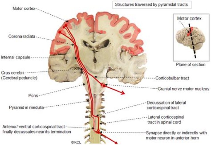
The corticobulbar tract is a two-neuron path which unites the cerebral cortex with the cranial nerve nuclei in the brainstem involved in motor functions (apart from the oculomotor nerve)..
In this regard, what is the difference between corticospinal and Corticobulbar tracts?
The corticobulbar tract conducts impulses from the brain to the cranial nerves. The corticospinal tract conducts impulses from the brain to the spinal cord. It is made up of a lateral and anterior tract. The corticospinal tract is involved in voluntary movement.
where does the Corticobulbar tract originate? The corticobulbar tract originates in the primary motor cortex of the frontal lobe, just superior to the lateral fissure and rostral to the central sulcus in the precentral gyrus (Brodmann area 4).
One may also ask, what is the corticospinal tract?
The corticospinal tract is a white matter motor pathway starting at the cerebral cortex that terminates on lower motor neurons and interneurons in the spinal cord, controlling movements of the limbs and trunk. The corticospinal tract is one of the pyramidal tracts, the other being the corticobulbar tract.
Does the Corticobulbar tract cross?
The corticobulbar tracts arise from the lateral aspect of the primary motor cortex. They receive the same inputs as the corticospinal tracts. The fibres converge and pass through the internal capsule to the brainstem.
Related Question Answers
Why do nerves cross over in the brain?
OBJECTIVE: In the chordate and vertebrate central nervous system, sensory and motor nerve tracts cross from one side to the other as they connect the brain with sensory receptors and motor neurons. These "decussations," crossings in the form of an X, relate each side of the brain to the opposite side of the body.Where does Corticobulbar end?
Cells of the cerebral hemispheres project varying nerve fibres that travels through the brainstem and terminate in the medulla oblongata (corticobulbar or corticonuclear tract) and in the spinal cord (corticospinal or pyramidal tract).Where do Corticobulbar tracts Decussate?
Approximately 85% of these primary motor axons decussate at the level of the medulla to cross to the contralateral spinal cord to enter the lateral corticospinal tracts.What does damage to the corticospinal tract cause?
Injuries to the lateral corticospinal tract results in ipsilateral paralysis (inability to move), paresis (decreased motor strength), and hypertonia (increased tone) for muscles innervated caudal to the level of injury. [2] The lateral corticospinal tract can suffer damage in a variety of ways.How does Decussation occur?
Decussation: when fibers cross from one side of a structure to the other. For example, motor fibers that travel in the corticospinal tract originate in the cerebral cortex and travel down to the body.How are spinal tracts named?
As the name suggests, the ascending tracts of the spinal cord ascend from the spinal cord and connect it to the brain. These tracts are named based on their origin and termination. They are found running along the dorsal, lateral, and ventral columns of the white matter.Is descending tracts sensory or motor?
Tracts descending to the spinal cord are involved with voluntary motor function, muscle tone, reflexes and equilibrium, visceral innervation, and modulation of ascending sensory signals. The largest, the corticospinal tract, originates in broad regions of the cerebral cortex.Where are interneurons located?
Interneurons (also known as association neurons) are neurons that are found exclusively in the central nervous system. That means that they are found in the brain and spinal cord and not in the peripheral segments of the nervous system.What is Decussation?
Definition of decussation. 1 : the action of crossing (as of nerve fibers) especially in the form of an X. 2 : a crossed tract of nerve fibers passing between centers on opposite sides of the nervous system.What is the function of the lateral corticospinal tract?
Function. Axons in the lateral corticospinal tract leave out of the tract and into the anterior horns of the spinal cord. It controls fine movement of ipsilateral limbs (albeit contralateral to the corresponding motor cortex) as it lies distal to the pyramidal decussation.Is the corticospinal tract motor or sensory?
Motor: The corticospinal tracts send motor information from the cortex to the spinal cord as the name suggests. Sensory: The anterolateral (or spinothalamic) tracts and dorsal (or posterior) column pathways bring sensory input from the spinal cord to the brain by way of the brainstem.Which tract crosses over in the brainstem to its opposite side?
The axons of the tract cells cross over (decussate) to the other side of the spinal cord via the anterior white commissure, and to the anterolateral corner of the spinal cord (hence the spinothalamic tract being part of the anterolateral system).What does the anterior Spinothalamic tract do?
The anterior spinothalamic tract, also known as the ventral spinothalamic fasciculus, is an ascending pathway located anteriorly within the spinal cord, primarily responsible for transmitting coarse touch and pressure.What is the difference between an upper and lower motor neuron?
The nerves that send messages between the cerebral cortex and the spine are called upper motor neurons, and those that relay messages from the spine to the muscles are called lower motor neurons.What does the pyramidal tract control?
These are called as pyramidal tracts as they crossover at the level of the pyramids in the medulla. They are collections of upper motor neuron fibers which go to the spinal cord (corticospinal) or the brainstem (corticobulbar) and control the motor function of the body.Where is the sensory Decussation?
The sensory decussation or decussation of the lemniscus is a decussation or cross over of axons from the gracile nucleus and cuneate nucleus. The fibres of this decussation are called the internal arcuate fibres and are found at the superior aspect of the closed medulla superior to the motor decussation.What contains cell bodies of upper motor neurons?
The cell bodies of these neurons are located within the ventral horns of the spinal cord and within brainstem motor nuclei. Upper motor neurons, as defined clinically, are cortical neurons that innervate lower motor neurons (either directly or via local interneurons).Are all cranial nerves lower motor neurons?
Lower motor neurons (LMNs) are motor neurons located in either the anterior grey column, anterior nerve roots (spinal lower motor neurons) or the cranial nerve nuclei of the brainstem and cranial nerves with motor function (cranial nerve lower motor neurons).What kind of fibers are upper motor neurons?
The upper motor neuron descends in the spinal cord to the level of the appropriate spinal nerve root. At this point, the upper motor neuron synapses with the lower motor neuron, each of whose axons innervate a fiber of skeletal muscle.