What happens if the Abducens nerve is damaged?
.
Likewise, what cranial nerve is affected by an Abducens nerve injury?
The abducens nerve has the longest intracranialcourse of any cranial nerve. It is primarily responsiblefor ipsilateral eye abduction. Abducens nerve palsyresults in an inability of the abducens nerve to transmitsignals to the lateral rectus, resulting in an inability to abductthe eye and horizontal diplopia.
One may also ask, how do you test Abducens nerve? The abducens nerve is examined in conjunctionwith the oculomotor and trochlear nerves by testingthe movements of the eye. The patient is asked to follow a pointwith their eyes (commonly the tip of a pen) without moving theirhead.
Beside above, what does the Abducens nerve control?
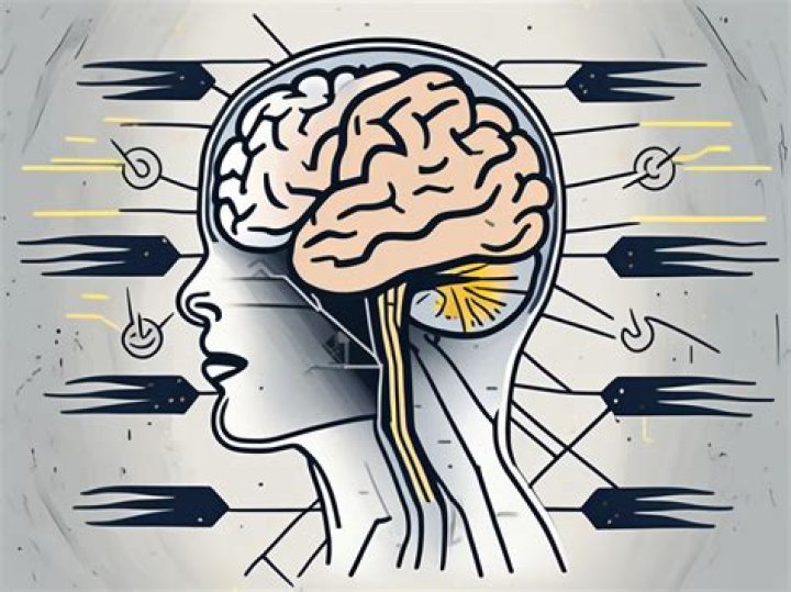
The abducens nerve is a nerve thatcontrols the movement of the lateral rectus muscle inhumans, responsible for outward gaze. It is also known as theabducent nerve, the sixth cranial nerve, sixthnerve, or simply CNVI. It is a somatic efferentnerve.
Is there a cure for sixth nerve palsy?
In some cases, sixth nerve palsy will disappearwithout treatment. If inflammation of the sixth nerveis suspected, medications called corticosteroids may be used. Untilthe nerve heals, wearing an eye patch can help with doublevision. Prism spectacles can also help to realigneyesight.
Related Question AnswersWhere is the 6th nerve?
Sixth nerve palsy is a disorder that affects eyemovement. It's caused by damage to the sixth cranial nerve.The primary function of the sixth cranial nerve is to sendsignals to your lateral rectus muscle. This small muscle is locatedon the outer side in your eye.What happens if the facial nerve is damaged?
When herpes zoster affects the facialnerve, the condition is called Ramsay Hunt syndrome. Toxins cancause damage to the facial nerve like alcohol; thedisease happens in chronic alcoholism. It involves therelatively quick onset of complete facial paralysis andinability to close the eye on one side of the face.Is 6th nerve palsy serious?
Isolated sixth nerve palsy itself does notusually cause complications. But many of the possible causes ofsixth nerve palsy may have complications. Like anyprocedure, surgery for sixth nerve palsy carries a risk forcomplications.What is CN palsy?
Cranial nerve palsy is characterized by adecreased or complete loss of function of one or more cranialnerves. The etiology may be congenital or acquired. Multiplecranial neuropathies are common, particularly in lesions arisingfrom tumors, trauma, impaired blood flow, andinfections.What is the 6th nerve palsy?
Sixth nerve palsy, or abducens nervepalsy, is a disorder associated with dysfunction of cranialnerve VI (the abducens nerve), which is responsiblefor causing contraction of the lateral rectus muscle to abduct(i.e., turn out) the eye.How do you test cranial nerve 6?
Cranial nerve VI controls eye movement to thesides. Ask the patient to look toward each ear. Then have himfollow your fingers through the six cardinal fields of gaze.Here's another easy technique you can use: With your finger, make abig X in the air and then draw a horizontal line acrossit.What are false localising signs?
False localising signs. Neurological signshave been described as "false localising" if they reflectdysfunction distant or remote from the expected anatomical locus ofpathology, hence challenging the traditional clinicoanatomicalcorrelation paradigm on which neurological examination isbased.Which cranial nerve is responsible for blurred?
The four cranial nerves involved in visionand movement of the eyes are the optic (I) nerve,oculomotor (III) nerve, trochlear (IV) nerve and theabducen (VI) nerve. The optic nerve is the sensorynerve for vision. It transmits information from theeyes to the brain.What does the Trochlear nerve do?
The trochlear nerve, also called the fourthcranial nerve or CN IV, is a motor nerve (a somaticefferent nerve) that innervates only a single muscle: thesuperior oblique muscle of the eye, which operates through thepulley-like trochlea.Where is the accessory nerve located?
They are branchiomotor in function and innervate thesternocleidomastoid and trapezius muscles in the neck and back. Thecranial root of the accessory nerve originates from cellslocated in the caudal medulla. They are found in the nucleusambiguus and leave the brainstem with the fibers of the vagusnerve.How do you test for trigeminal neuralgia?
Test for motor abnormalities as follows:- Observe the skin over the temporal masseter muscles.
- Ask the patient to clench his or her jaws.
- Observe for deviation of the tip of the mandible as the jawsare opened.
- Ask the patient to move the jaw from side to side against theresistance of your palm.