How long does a head ultrasound take?

.
Similarly, you may ask, what does an ultrasound of the head show?
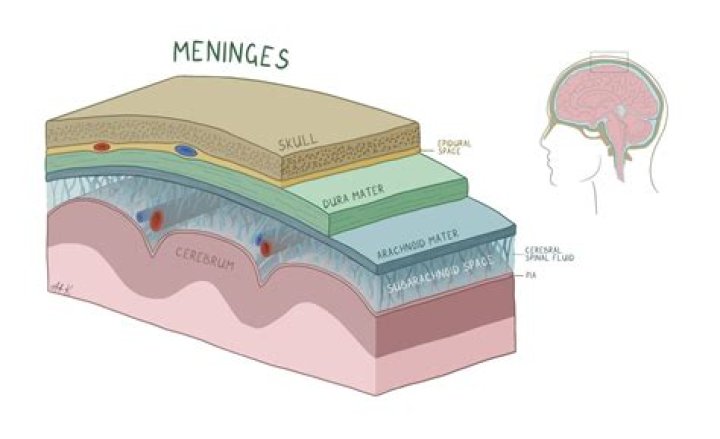
Ultrasound imaging of the head uses sound waves to produce pictures of the brain and cerebrospinal fluid. It is most commonly performed on infants, whose skulls have not completely formed. A transcranial Doppler ultrasound evaluates blood flow in the brain's major arteries.
how long does an ultrasound take to get results? How long will it take for my doctor to receive the results of my ultrasound exam? One of our board-certified radiologists will review and interpret your ultrasound results immediately. Your doctor will receive a written report and hardcopy images within 24 hours.
Additionally, can brain damage be seen on ultrasound?
Ultrasound 'may damage baby's brain' Ultrasound scans for pregnant women could cause brain damage in their unborn babies, research warns. Doctors found that men born to mothers who underwent scanning were more likely to show signs of subtle brain damage.
How long does a head and neck ultrasound take?
The ultrasound probe is moved gently over the skin. The images are displayed on a monitor and are viewed by our consultant radiologist (a specialist in x-rays). The ultrasound scan does not usually take more than 30 minutes. Any gel which might get onto your clothes will wash off easily.
Related Question AnswersCan you see cancer on a ultrasound?
Ultrasounds Often Fail to Detect Cancer Therefore, an ultrasound can't tell a cancerous tumour from a benign tumour. "Sometimes imaging tests can show something that looks like cancer, but further tests (such as a biopsy) show that it's not cancer."How do they do a head ultrasound?
A head ultrasound is a safe and painless test that uses sound waves to make images of the brain. During the examination, an ultrasound machine sends sound waves into the head and images are recorded on a computer. The fontanel provides an opening for the sound waves to get through and reach the brain.What does fluid look like on an ultrasound?
Fluid presents has an anechoic appearance on ultrasound, and can be confirmed with dynamic interrogation as it should respond to pressure. You can see here the anechoic or black appearance of fluid within the superficial infrapatellar bursa of the knee.What abnormalities can be detected on an ultrasound?
Ultrasound can detect some types of physical birth defects. Examples of physical birth defects that may be found at 19 - 20 weeks are most cases of spina bifida, some serious heart defects, some kidney problems, absence of part of a limb and some cases of cleft palate.What is a cranial scan?
A cranial CT scan is a diagnostic tool used to create detailed pictures of features inside your head, such as your skull, brain, paranasal sinuses, ventricles, and eye sockets. A cranial CT scan is known by a variety of names as well, including brain scan, head scan, skull scan, and sinus scan.Why do an ultrasound of the neck?
Carotid (kuh-ROT-id) ultrasound is a safe, painless procedure that uses sound waves to examine the blood flow through the carotid arteries. Your two carotid arteries are located on each side of your neck. They deliver blood from your heart to your brain.How long does a vascular ultrasound take?
This ultrasound examination is usually completed within 30 to 45 minutes. Occasionally, complex examinations may take longer.What is ultrasound of head and neck?
Diagnostic ultrasound in the head and neck region. Ultrasound is the first line imaging modality for suspected salivary gland tumours and is useful in evaluating superficial neck swellings, such as lymph node disease, soft tissue cysts and vascular malformations.What are the symptoms of brain damage?
Mild traumatic brain injury symptoms may include:- Inability to remember the cause of the injury or events that occurred immediately before or up to 24 hours after it happened.
- Confusion and disorientation.
- Difficulty remembering new information.
- Headache.
- Dizziness.
- Blurry vision.
- Nausea and vomiting.