Does the Tectorial membrane vibrate
The more rigid a tectorial membrane is, the higher the frequency at which it can vibrate. Thus, the flexible end of the membrane—which should respond to low-frequency vibration— is found near the hair cells that transmit low frequencies, and the rigid end near hair cells that transmit high ones.
What does the tectorial membrane do?
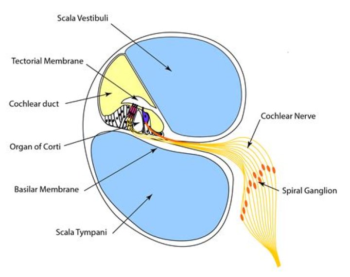
The tectorial membrane (TM) is an extracellular connective tissue that covers the mechanically-sensitive hair bundles of the sensory receptor cells in the inner ear. It occupies a strategic position, playing a key role in transforming sound to mechanical stimulation.
What causes the tectorial membrane to move?
A tectorial (roof) membrane is held in place by a hinge-like mechanism on the side of the Organ of Corti and floats above the hair cells. As the basilar and tectorial membranes move up and down with the traveling wave, the hinge mechanism causes the tectorial membrane to move laterally over the hair cells.
What membrane vibrates in the ear?
The Outer Ear The sound waves then travel toward a flexible, oval membrane at the end of the ear canal called the eardrum, or tympanic membrane. Sound waves cause the eardrum to vibrate.What is the role of the tectorial membrane in sound detection?
When sound stimulates the stereocilia on the sensory cells in the hearing organ, Ca2+ ions flow through mechanically gated ion channels. … Hence, the tectorial membrane contributes to control of hearing sensitivity by influencing the ionic environment around the stereocilia.
What are alar ligaments?
In anatomy, the alar ligaments are ligaments which connect the dens (a bony protrusion on the second cervical vertebra) to tubercles on the medial side of the occipital condyle.
Is oval window inner ear?
The oval window, also known as the fenestra ovalis, is a connective tissue membrane located at the end of the middle ear and the beginning of the inner ear.
Why does my eardrum keep vibrating?
Fluttering in the ear is an annoying symptom that can affect a person’s quality of life. People may have difficulty hearing and focusing. Doctors suggest that fluttering in the ear is a type of tinnitus called MEM, which is caused by jerky movements of the muscles in the middle ear.What vibrates directly after the tympanic membrane vibrates?
The eardrum vibrates. The vibrations are then passed to 3 tiny bones in the middle ear called the ossicles. The ossicles amplify the sound. They send the sound waves to the inner ear and into the fluid-filled hearing organ (cochlea).
What is the first structure that vibrates in response to sound waves entering the external ear?Sound waves enter the outer ear and travel through a narrow passageway called the ear canal, which leads to the eardrum. The eardrum vibrates from the incoming sound waves and sends these vibrations to three tiny bones in the middle ear. These bones are called the malleus, incus, and stapes.
Article first time published onWhat directly causes vibrations of the basilar membrane of the organ of Corti?
The motion of the stapes against the oval window sets up waves in the fluids of the cochlea, causing the basilar membrane to vibrate. This stimulates the sensory cells of the organ of Corti, atop the basilar membrane, to send nerve impulses to the brain.
Which structure converts vibrations into nerve impulses?
The cochlea is filled with a fluid that flows in response to the vibrations from the oval window. As the fluid moves, 25,000 nerve endings are set into motion. These nerve endings transform the vibrations into electrical impulses that then progress along the eighth cranial nerve (auditory nerve) to the brain.
What happens to the hair cells when the basilar membrane vibrates?
When sound-induced basilar membrane vibrations deflect hair bundles of the outer hair cells, mechanoelectrical transduction of these cells generates the receptor potential (Dallos et al., 1982; Russell and Sellick, 1983).
What dissipates vibrations within the cochlea?
The mechanical vibrations of the stapes footplate at the oval window creates pressure waves in the perilymph of the scala vestibuli of the cochlea. These waves move around the tip of the cochlea through the helicotrema into the scala tympani and dissipate as they hit the round window.
What happens when stereocilia bend?
Bending the stereocilia toward the kinocilium depolarizes the cell and results in increased afferent activity. Bending the stereocilia away from the kinocilium hyperpolarizes the cell and results in a decrease in afferent activity. The semicircular ducts work in pairs to detect head movements (angular acceleration).
What are stereocilia and kinocilium?
one relatively long hair (kinocilium) and about 50 shorter ones (stereocilia). The kinocilium is inserted eccentrically on top of the sense cell; the stereocilia are arranged in parallel rows. In about half of the hair cells of a neuromast, the kinocilium is found on one (and the same) side…
Are ears connected to each other?
These bones are connected to each other. The last in the group, stapes, also makes contact with the inner ear. The air space of the middle ear connects to the back of the nose by the Eustachian tube, a narrow tube which can let air in or out of the space.
How far down is your eardrum?
The eardrum is located about 1.5 cm inside the skull at the end of the external auditory canal. The canal is rigid (surrounded by bone and cartilage), but it is not straight.
What is auricle ear?
The medical term for the outer ear is the auricle or pinna. The outer ear is made up of cartilage and skin. There are three different parts to the outer ear; the tragus, helix and the lobule. EAR CANAL. The ear canal starts at the outer ear and ends at the ear drum.
What is Atlas and axis?
The atlas and axis vertebrae are the two most superior bones in the vertebral column, and they are part of the seven cervical vertebrae. The atlas is the top-most bone, sitting just below the skull; it is followed by the axis. Together, they support the skull, facilitate neck movement, and protect the spinal cord.
Where does alar ligaments attach?
The alar ligaments join the lateral margins of the sloping upper posterior margin of the dens of C2 to the lateral margins of the foramen magnum (adjacent to the occipital condyles) and lie on either side of the apical ligament. They may be oblique or vertical and are thickest at the occipital attachment.
What is Jefferson fracture?
A Jefferson fracture is a bone fracture of the vertebra C1. The vertebra C1 is a bony ring, with two wedge-shaped lateral masses, connected by relatively thin anterior and posterior arches and a transverse ligament. The lateral mass on vertebra C1, who is taller, is directed laterally.
Is sound turned into a nerve message?
The ear is divided into three regions: the outer ear, the middle ear, and the inner ear. While the first two sections collect and transmit sound as waves/vibrations, the inner ear, comprising the cochlea and semicircular canals is responsible for converting that physical energy into electrical energy (nerve impulses).
Which is the last structure to vibrate in this sequence?
So the last structure to vibrate in this sequence is oval window. Arrange the following in the order of reception and transmission of the sound wave from the eardrum. Cochlear nerve, external auditory canal, drum, stapes, incus, malleus, cochlea. Malleus, Iris, Stapes, Incus.
Which will start to vibrate the vibration will become amplified as it moves through the ossicles in the order of?
The vibration will become amplified as it moves through the middle ear’s bones, the ossicles in the order of HAMMER, ANVIL, and STIRRUP. Thereafter, the sound vibration goes into the inner ear’s oval window and reaches the COCHLEA, a fluid snail shaped object.
Why is my ear making bubbling sounds?
The bottom line. Sometimes you may experience crackling or popping in your ears. This is often described as a “Rice Krispie”-like sound. Crackling in the ears can be caused by several different conditions, such as eustachian tube dysfunction, acute otitis media, or the buildup of earwax.
Will ear fluttering go away?
Tensor Tympani Spasms are a condition which causes “shotgun” or “fluttering” sounds in the ear. The tinnitus may only last for a brief moment, or it can last as long as a few days. Many people who experience Tensor Tympani Spasms assume that these sensations are normal.
Which structure transmits sound vibrations to the perilymph of the cochlea which structure transmits sound vibrations to the perilymph of the cochlea?
The stapes does not move in and out but rocks back and forth about the lower pole of its footplate, which impinges on the membrane covering the oval window in the bony plate of the inner ear. The action of the stapes transmits the sound waves to the perilymph of the vestibule and the scala vestibuli.
Why is it important for sound vibrations to be amplified as they pass through the ear?
Why is it important for sound vibrations to be amplified as they pass through the ear? More force is needed to create waves in fluid. … How do sound waves ultimately result in the production of receptor potentials? Hair cells in the cochlea vibrate, causing ion channels to open in their membrane.
Which of the following moves in response to the vibration of the stapes?
Sound waves travel along the auditory canal and strike the tympanic membrane, causing it to vibrate. This vibration results in movement of the three ossicles. As the ossicles move, the stapes presses into a thin membrane of the cochlea known as the oval window.
How does the basilar membrane distinguish different frequency vibrations?
The BM presents the first level of frequency analysis in the cochlea because of its changing stiffness and mass from base to apex. High-frequency sound produces maximal BM movement at the “base” of the cochlea (near the stapes) whereas low-frequency sound also activates the apical parts of the BM.