Does bulbar ALS show on EMG?
Does bulbar ALS show on EMG?
Electromyography (EMG), particularly measurements of the tongue or sternocleidomastoid, aids in the diagnosis of amyotrophic lateral sclerosis (ALS) and may be used to identify lower motor neuron lesions in the bulbar region. Abnormal trapezius EMG recordings were recently shown to be useful in diagnosing ALS.
Does bulbar ALS show up on MRI?
Conclusions: Imaging features on MRI can aide in the diagnosis of ALS, in addition to ruling out alternate pathology. These changes can be well delineated and observed in the subset population of bulbar onset ALS.
What test is used to confirm ALS?
Electromyography: EMG is one of the most important tests used to diagnose ALS. Small electric shocks are sent through your nerves. Your doctor measures how fast they conduct electricity and whether they’re damaged. A second part of the test also checks the electrical activity of your muscles.
What are bulbar ALS symptoms?
Symptoms Affecting Speech Harsh, hoarse or strained voice. Breathy speech pattern. Poor articulation. Decrease in range of pitch and loudness of voice.
Do I have bulbar onset ALS?
Although progression is variable by case, Bulbar Onset ALS tends to have a faster progression than Limb Onset cases. Early symptoms include slurred speech, difficulty chewing and swallowing, excessive choking and weakness or twitching in the muscles of the face, jaw, throat and voice box, particularly the tongue.
How do you rule out bulbar ALS?
Doctors use physical examination to assess swallowing, lip and tongue strength and speech tests in addition to other neurological tests, to diagnose Bulbar Onset ALS.
What mimics bulbar ALS?
On the other hand, most studies show that bulbar-onset ALS displays a female predominance. In contrast to other neurodegenerative disorders, the risk of developing ALS peaks between the ages of 50 and 75 years, and declines thereafter.
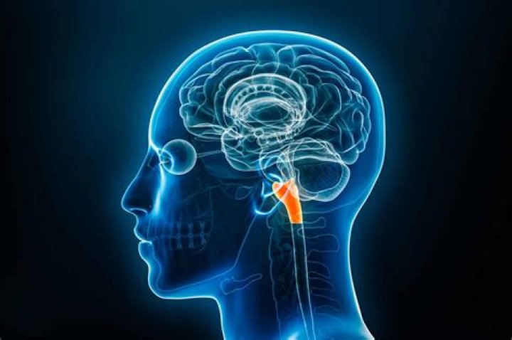
What is a bulbar palsy?
5.6. A bulbar palsy refers to disease affecting the glossopharyngeal, vagus, accessory and hypoglossal nerves and is due to lower motor neuron pathology. Typically, patients with a bulbar palsy present with signs and symptoms of the cranial nerves affected as mentioned.
How often is ALS misdiagnosed?
Misdiagnosis works both ways: 10-15% of cases are false positive, which means patients are told they have ALS, but their symptoms end up being due to some other condition. Nearly 40% of patients are false-negative, meaning they are diagnosed as having some other condition before ALS is confirmed.
How quickly does bulbar ALS progress?
The median time to symptomatic progression beyond the bulbar region was approximately 1 year, with equal proportions progressing to the upper or lower limbs. The median interval from onset to anarthria was 18 months, and to loss of ambulation 22 months.
How aggressive is bulbar ALS?
In the bulbar-onset group, the damage in the gray matter was associated with ALSFRS-R scores, and the forced vital capacity was linked to damage in deeper brain structures. The findings support earlier studies and clinical observations that bulbar-onset patients have a more aggressive disease.